
Interactive Anatomy Visualization App
Medical UX & 3D Design Case Study
This project involved the design and development of an interactive medical application for visualizing the musculoskeletal anatomy of the arm. Combining 3D modeling, real-time interaction, and user-centered design, the tool supports medical and kinesiology students in understanding complex anatomical relationships that are difficult to grasp through static imagery.
Developed as part of a Master’s in Biomedical Communications, the project focuses on translating dense anatomical information into an intuitive, exploratory learning experience.
OVERVIEW
Understanding musculoskeletal relationships in the arm is challenging due to overlapping structures, depth, and movement. Traditional learning tools such as textbooks and static diagrams often fail to convey these spatial relationships effectively.
This project addresses that gap by creating an interactive anatomy application that allows users to explore muscles, bones, and their functional connections in a dynamic 3D environment.
Key Contributions:
UX design and interaction modeling
3D anatomical modeling and optimization
Interface design for educational use
Integration into a real-time interactive environment
Award of Excellence 2017
Association of Medical Illustrators

DESIGN PROCESS (UX & WIREFRAMING)
The interaction design focused on simplicity and clarity. Early wireframes explored how users could navigate anatomical layers, isolate structures, and understand functional relationships without becoming overwhelmed.
Key considerations included:
Minimizing cognitive load
Creating intuitive controls for non-technical users
Allowing progressive exploration of complexity
Wireframing and iterative testing informed the layout, navigation patterns, and interaction logic of the final application.
3D MODELING & TECHNICAL DEVELOPMENT
High-resolution anatomical models were developed and optimized for real-time performance. Particular attention was given to balancing anatomical accuracy with usability and responsiveness.
Key aspects of the modeling process:
Sculpting and refining anatomical structures
Retopology and optimization for real-time rendering
Material and shading adjustments for clarity and readability
The models were integrated into an interactive environment, enabling users to manipulate and explore structures dynamically.
INTERACTION & IMPLEMENTATION
The application was built within a real-time environment, where interaction logic was developed to allow users to isolate, manipulate, and explore anatomical structures.
Implementation focused on:
Responsive interaction design
Clear visual feedback
Maintaining performance with complex 3D assets
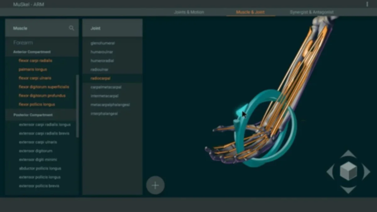
OUTCOME
The final application enables intuitive exploration of musculoskeletal anatomy through direct manipulation of individual muscles and joints. By interacting with structures in real time, users can better understand spatial relationships and functional behavior that are difficult to grasp through static imagery.
The project was recognized with an Award of Excellence (2017) from the Association of Medical Illustrators.
Developed as part of a Master’s in Biomedical Communications at the University of Toronto, under the supervision of Michael Corrin and Judi Laprade.